
Software
For over 25 years, Brainsight has been setting the standard for functionality and ease of use for TMS and veterinary neuronavigation and we are bringing this to bear for fNIRS. This approach gives you the capability of mapping all your fNIRS (as well as your TMS, EEG and EMG) to either standard brain template (e.g. MNI brain), or to the subject-specific MRI. You can decide how much precision you need and consequently how much effort is acceptable.
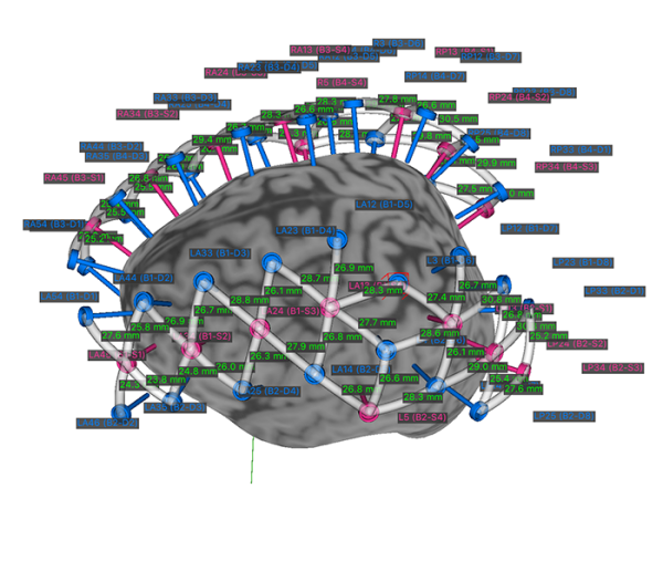
Our montage manager enables you to define your common fNIRS and EEG configurations for quick recall at the start of a fNIRS session. The locations of the optodes can be defined in a similar way as is done in EEG. The initial locations can be defined in a standard 3D coordinate (MNI space) and automatically mapped to the individual’s MR images. The tracked pointer can be used to further refine the actual location once the optodes are placed on the head at the start of the fNIRS session.
Define your caps
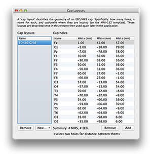
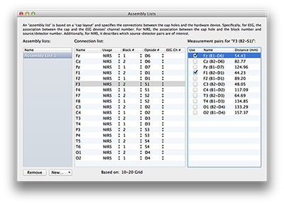
If you perform the same montage of optodes (and/or EEG electrodes) on multiple subjects using a cap to hold the optodes, you can define the cap once using our cap manager. The definition can contain the 3D locations of each optode in the standard MNI space as well as the intended purpose of the optode, be it source or detector (as well as EEG electrode). This cap and assembly list definition can be instantly applied at acquisition time for the subject either to the standard template brain (if no subject MRI is available), or warped to the subject specific MRI. If maximum precision is needed, you can use the 3D tracked pointer to record the exact location of each optode and Brainsight maps it back to the subject specific MR space, or to MNI space (or both if desired).
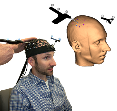
Apply the cap
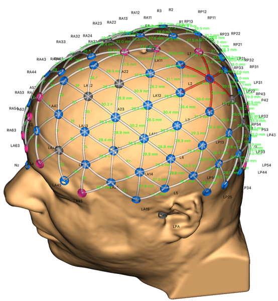
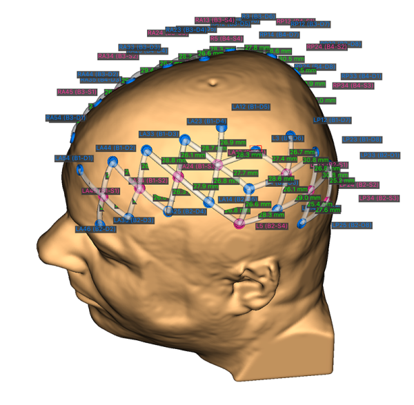
You can use a full cap, or a patch that optimizes coverage over your target area.
The locations of the optodes/electrodes can be pre-defined in the MNI standard coordinate space, or you can use the neuronavigator to accurately digitize the locations and map them to the subject-specific MRI.
Acquire your data
Once you are ready, performing the experiment can be done knowing your optodes are well placed and the actual locations can be exported and used in data analysis.