For research applications, Brainsight can be used for a wide variety of procedures, including cannula and needle placement as well as chamber positioning for electrophysiology. Brainsight® Vet brings the freedom and flexibility of frameless stereotaxy with the accuracy of frame based systems using our novel fiducial marker technology.
Fiducial Markers
Our unique fiducial marker array system provides a rigid, unambiguous set of landmarks that allows accurate (about 1 mm) co-registration of the subject to the images at surgery. The array is mounted to either a fixation post or small dedicated post during imaging and surgery. This means that you do not have to perform the surgery immediately after imaging and you can re-use the images for multiple procedures. This saves time and money (lower scanner costs), is easier for the subject (less anaesthesia), and by removing the need for a stereotactic frame, more comfortable. The scanning is simpler and you can take advantage of a wide array of MRI coils to obtain even better images.

Surgical Planning: Needle Placement
Brainsight includes sophisticated 3D image reconstruction tools to allow you to explore the brain prior to surgery. In the case of needle or single electrode placement, you can decide on your target(s) without the time pressure of performing the surgery immediately after imaging. Without the frame, you have the freedom to pick any approach to your target that avoids critical structures without having to perform complex calculations. Brainsight stores the paths to target and guides you in surgery using our Freeguide™ tool platform and articulated arm.
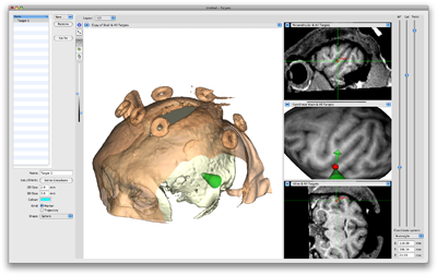
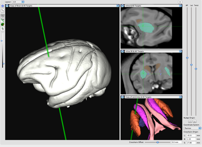
Segment critical structures using the ROI tool and visualize them in 2D or 3D. Co-register the animal’s brain to selected atlases to correlate anatomical targets.
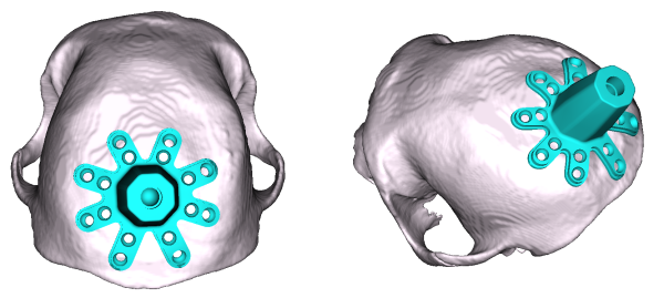
Surgical Planning: Electrophysiology Chamber Placement
One of the best uses of Brainsight is to plan the optimal location for your electrophysiology chambers and grids. Simulate the type of chamber (square, round) and virtually place it anywhere on the skull. You can view the trajectory of each needle track of the grid into the brain to ensure that you can reach your targets. Once implanted, you can record the final chamber location and use the software to project the actual grid location onto the MR images. This allows you to find the best grid tracks and depth to target to reduce the time it takes to place your electrodes to record from the right cells.
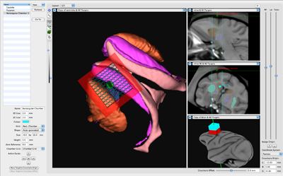
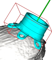
Brainsight supports industry standard CAD files (STL, polygonal DXF) so you can simulate the placement of the chamber on the skull prior to surgery. Plan out the surgery to the smallest detail, down to the last screw. All this is later displayed in surgery to ensure that you follow the plan. Record the final location in surgery for use later to simulate the insertion of each electrode. Determine the optimal grid selection and depth to target before the first recording session to simplify data gathering.
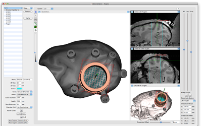
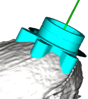
Surgical Planning: Custom Implant Design
Take impant design to the next level. Working with our state of the art machine shop equipment and staff, you can use the CAD representation of your implant, the planned location and the 3D rendering of the skull to sculpt a custom implant that fits perfectly on the bone. This reduces or eliminates the need for bone cement, and improves the overall chances of a sucessful implant.
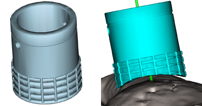
We can further customize the implant by adding fixation screw mounting points. The result requires little or no cranial cement to fix, reducing the overall size of the implant.